Dental Disease and Oral Pain – Case Study #2
This case report deals with an issue in kitty cats that these days is less often overlooked. To this point there are have no answers as to the ultimate cause or strategy for prevention. This serious feline dental disease was the subject of one of AVC’s earliest case studies posted on the website and is certainly worth visiting again.
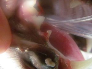
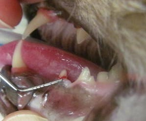
The photos of Max’s teeth are quite graphic as they show extreme erosion of the involved teeth, a condition known by several monikers: Feline Oral Resorptive Lesions, Feline Odontoclastic Lesions and “Neck” Lesions.
Regardless of the terminology, resorptive lesions are the result of a cell called the odontoclast becoming active is removing enamel from the teeth. This results in a structurally weakened and with exposure of the dentin layer which is the innervated area of the tooth. Consequently, affected teeth are quite painful. It is thought that the odontoclast was at one time an Odontoblast which is the cell responsible for laying down tooth enamel in the first place. (“Blast cells make stuff while “Clast” cells remove stuff.) If that is the case, some smart person will have to figure out what causes the change in format from Blast to Clast. The catch-all phrase for things we don’t understand about cat disease process is “It is probably immune mediated.”
That may sound intuitive, but it actually means exactly this, “DUNNO!”
Symptoms are frequently quite subtle and may include gradual weight loss, hiding, skittish-ness, vocalizing, scratching at the face or ears, tacky hair coat or unthrifty overall appearance and change in otherwise friendly behavior. Many of our kitty families have reported in retrospect such things as swallowing food whole, pitching the head back when eating and eating tiny portions, all related to the kitty trying to avoid using those painful teeth.
Resolution of lesions such as Max exhibited requires extraction of the damaged, painful teeth, following which most kitties flourish. Max certainly did.
These photos show severe FORLs of lower right and left pre-molars respectively. The left hand photo shows the loss of the entire caudal portion of the tooth for the gum line (neck) toward the crown. The right hand photo demonstrates a typical finding with FORLs in that inflammatory gingival tissue, the red ring around the neck of the tooth is growing upward in an attempt to cover the damaged area of the tooth.
Latest Blog
Mar
09, 2023Feb
23, 2023Jan
05, 2023Mar
26, 2024Mar
26, 2024Mar
26, 2024Mar
23, 2023